The jam board linked to these images contains super-simple graphics to prompt conversation about the differences between EEG and fMRI, add stickies, draw on it, whatever you like, once you click you can make your own editable copy. Just scroll through the slides to find the activity you want.
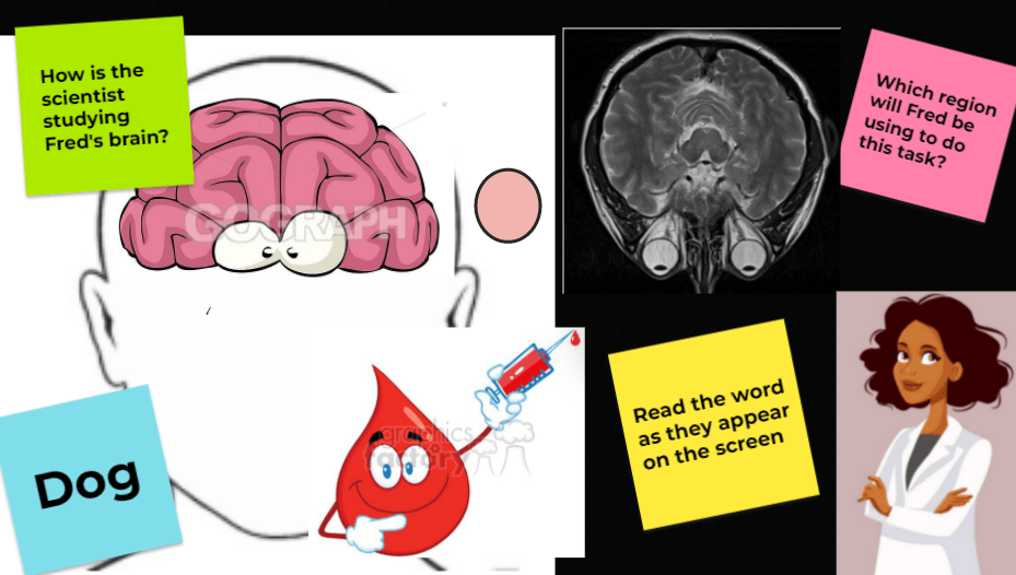
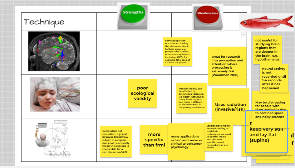
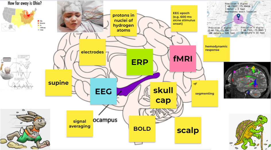
Discussing fMRI: Revise Broca’s area, (place the blob in the correct hemisphere, name the lobe), use the blood droplet to discuss the fact that more oxygenated blood will be required in this location and how the fMRI measures this (refers to BOLD contrast) show the area ‘lighting up’ on the scan (use warm colours for active areas and cool colours for inactive areas). Lots and lots of sticky notes to sort into strengths and weaknesses of fMRI, EEG and ERP. Change the colour of the stickies (green for strength and orange for weakness). There is one red herring which is not true of any of the techniques, place the fish on the red herring!Revising specialist terminology associated with fMRI, EEG and ERP, students can add more stickies if they can think of more specialist terms! Change the sticky colour to match the technique that they are related to. Place the tortoise and the hare on the techniques that have excellent temporal resolution (hare) and poor temporal resolution (tortoise). Place the accurate map on the technique with strong spatial resolution and the poor map on the technique with poor spatial resolution. Join the related words up using the pens (like they are in a neural network) discuss how the concepts are stored within semantic webs of associative memory, firing in one area of the network should trigger therm to think of all the related words.